Connect With Us
Blogs

Sunday, 22 June 2025 00:00
Ankle Fracture? Don’t Wait for Treatment


If you're suffering from an ankle fracture, professional attention shouldn't wait. Fractures may worsen over time and can make completing everyday activities incredibly difficult. See your specialist for treatment and guidance through the healing process.
Published in Blog
Tagged under
Tuesday, 17 June 2025 00:00
Flat Feet in Women

Flat feet can have a significant impact on posture, movement, and stability, especially in females. The arch of the foot plays a key role in absorbing shock and distributing weight. When the arch collapses, it can affect balance, reduce agility, and place additional strain on core muscles, leading to fatigue and instability. Symptoms may include foot pain, swelling, and difficulty with physical activities. Risk factors include genetics, pregnancy, and obesity. A podiatrist can evaluate foot structure, recommend supportive footwear, and assign exercises that improve strength and alignment. If you have flat feet and notice persistent foot discomfort or instability, it is suggested that you consult a podiatrist for help in restoring function and improving overall movement.
Flatfoot is a condition many people suffer from. If you have flat feet, contact Michael Schwartzman, DPM from Illinois and Indiana . Our doctor will treat your foot and ankle needs.
What Are Flat Feet?
Flatfoot is a condition in which the arch of the foot is depressed and the sole of the foot is almost completely in contact with the ground. About 20-30% of the population generally has flat feet because their arches never formed during growth.
Conditions & Problems:
Having flat feet makes it difficult to run or walk because of the stress placed on the ankles.
Alignment – The general alignment of your legs can be disrupted, because the ankles move inward which can cause major discomfort.
Knees – If you have complications with your knees, flat feet can be a contributor to arthritis in that area.
Symptoms
- Pain around the heel or arch area
- Trouble standing on the tip toe
- Swelling around the inside of the ankle
- Flat look to one or both feet
- Having your shoes feel uneven when worn
Treatment
If you are experiencing pain and stress on the foot you may weaken the posterior tibial tendon, which runs around the inside of the ankle.
If you have any questions, please feel free to contact our offices located in West Chicago, IL and Whiting, IN . We offer the newest diagnostic and treatment technologies for all your foot care needs.
Published in Blog
Tagged under
Tuesday, 17 June 2025 00:00
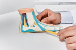
Flat Feet
Flatfoot is a foot condition in which the arch of the foot has either partially or totally dropped or has never developed. While it is common in babies and small children, it can become a problem for them in adulthood if the arch never forms. For adults, the development of flat feet can be brought upon by injury, as a result of pregnancy due to increased elasticity, or obesity. Those who have health concerns such as rheumatoid arthritis or diabetes may also be at greater risk for developing the condition.
If you suspect that you have flat feet, it is best to consult your podiatrist. Your foot doctor will examine the suspected foot and observe how it looks while you sit and stand. He or she may take an X-ray to determine how serious the condition is. Some common signs of flatfoot include toe drift, in which the toes and front part of the foot point outward, a short Achilles tendon, and a heel that tilts outwardly while the ankle tilts inward.
Once flatfoot has been diagnosed, your podiatrist may suggest one of several treatment options. Flat feet can be rigid, in which the feet appear to have no arch even when the person is not standing; or flexible, in which the person appears to have an arch while not standing, but once standing the arch disappears. Those with flexible flatfoot may be told to reduce any activities that cause pain and to avoid extended periods of walking or standing. Another suggestion may be weight loss, as excessive weight may be placing pressure on the arches
In few cases, if the condition is severe and all other methods have been exhausted surgery may be required. This is normally avoided, however, due to a lengthy recovery time and high cost.
Published in Featured
Tagged under
Tuesday, 10 June 2025 00:00
What Is Sesamoiditis?

Sesamoiditis is a painful condition involving inflammation of the sesamoid bones, two small bones located beneath the big toe joint. These bones help with movement and bear weight during walking and running. Sesamoiditis is usually caused by repetitive stress, high-impact activities, or wearing shoes that do not provide enough support. Symptoms include pain under the big toe, swelling, bruising, and difficulty bending or straightening the toe. Risk factors include participating in sports like running or dancing, having high arches, or spending long hours on hard surfaces. A podiatrist can diagnose sesamoiditis through examination and imaging, then recommend treatments such as rest, footwear changes, custom orthotics, and stretching exercises to reduce pain and promote healing. If you have pain in this part of your foot, it is suggested that you contact a podiatrist who can accurately diagnose the problem, and offer appropriate relief and treatment solutions.
Sesamoiditis is an unpleasant foot condition characterized by pain in the balls of the feet. If you think you’re struggling with sesamoiditis, contact Michael Schwartzman, DPM of Illinois and Indiana . Our doctor will treat your condition thoroughly and effectively.
Sesamoiditis
Sesamoiditis is a condition of the foot that affects the ball of the foot. It is more common in younger people than it is in older people. It can also occur with people who have begun a new exercise program, since their bodies are adjusting to the new physical regimen. Pain may also be caused by the inflammation of tendons surrounding the bones. It is important to seek treatment in its early stages because if you ignore the pain, this condition can lead to more serious problems such as severe irritation and bone fractures.
Causes of Sesamoiditis
- Sudden increase in activity
- Increase in physically strenuous movement without a proper warm up or build up
- Foot structure: those who have smaller, bonier feet or those with a high arch may be more susceptible
Treatment for sesamoiditis is non-invasive and simple. Doctors may recommend a strict rest period where the patient forgoes most physical activity. This will help give the patient time to heal their feet through limited activity. For serious cases, it is best to speak with your doctor to determine a treatment option that will help your specific needs.
If you have any questions, please feel free to contact our offices located in West Chicago, IL and Whiting, IN . We offer the newest diagnostic and treatment technologies for all your foot care needs.
Published in Blog
Tagged under
Tuesday, 10 June 2025 00:00
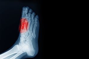
Sesamoiditis
Sesamoiditis is a condition in which the sesamoid bones in the forefoot become inflamed from physical activity. Sesamoid bones are bones that are not connected to other bones but are located in tendons or muscle. Two of these sesamoid bones are very small and located on the underside of the foot near the big toe. Athletes such as runners, baseball and football players, and dancers are likely to experience sesamoiditis. Those with high arched feet, flat feet, or runners who run on the ball of their foot are also prone to suffer from sesamoiditis.
Symptoms include pain or throbbing on the ball of the foot near the big toe. The pain generally starts with a mild throbbing but gradually builds up to shooting pain. Bruising, swelling, and redness are possible, but in most cases, these symptoms are not present. However, moving the big toe can result in pain and difficulty.
To conduct a diagnosis, the podiatrist will examine the ball of the foot and big toe. They will look for any outliers and check the movement of the toe. X-rays will be taken to rule out any other conditions and ensure that it is sesamoiditis.
Treatment for sesamoiditis is generally mild and includes rest, anti-inflammatory and pain medication, and ice treatments to deal with the swelling and pain. Orthotics may be needed with people who have flat or high arched feet to relieve pressure off the bones. In some cases the toe will be taped and immobilized to allow healing. The podiatrist may also decide to use a steroid injection to help with swelling as well. If you have sesamoiditis, you shouldn’t engage in any intensive activity, as it may inflame the area and worsen your pain. If the sesamoid bone has fractured, surgery may be required to remove the sesamoid bone.
If you are suffering from sesamoiditis or are experiencing symptoms similar to sesamoiditis, you should stop all physical activity that puts strain on the area. Furthermore you should see a podiatrist for a diagnosis to see if you have sesamoiditis.
Published in Featured
Tagged under
Tuesday, 03 June 2025 00:00
Common Types of Heel Spurs

Heel spurs are calcium deposits that form on the heel bone and can cause significant foot pain. The two most common types are posterior heel spurs, which develop at the back of the heel near the Achilles tendon, and plantar heel spurs, which form on the bottom of the heel and are often linked to plantar fasciitis. Causes include repeated strain on foot muscles and ligaments, long periods of standing, or improper footwear. Symptoms may involve sharp pain when standing or walking, especially after rest. Risk factors include obesity, flat feet, high arches, and increased physical activity. A podiatrist can diagnose heel spurs through imaging and provide personalized treatments. If you have persistent heel pain, it is suggested that you schedule an appointment with a podiatrist who can offer lasting relief and help prevent further complications.
Heel spurs can be incredibly painful and sometimes may make you unable to participate in physical activities. To get medical care for your heel spurs, contact Michael Schwartzman, DPM from Illinois and Indiana . Our doctor will do everything possible to treat your condition.
Heels Spurs
Heel spurs are formed by calcium deposits on the back of the foot where the heel is. This can also be caused by small fragments of bone breaking off one section of the foot, attaching onto the back of the foot. Heel spurs can also be bone growth on the back of the foot and may grow in the direction of the arch of the foot.
Older individuals usually suffer from heel spurs and pain sometimes intensifies with age. One of the main condition's spurs are related to is plantar fasciitis.
Pain
The pain associated with spurs is often because of weight placed on the feet. When someone is walking, their entire weight is concentrated on the feet. Bone spurs then have the tendency to affect other bones and tissues around the foot. As the pain continues, the feet will become tender and sensitive over time.
Treatments
There are many ways to treat heel spurs. If one is suffering from heel spurs in conjunction with pain, there are several methods for healing. Medication, surgery, and herbal care are some options.
If you have any questions, please feel free to contact our offices located in West Chicago, IL and Whiting, IN . We offer the newest diagnostic and treatment technologies for all your foot care needs.
Published in Blog
Tagged under
Tuesday, 03 June 2025 00:00
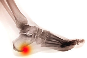
Heel Spurs
Heel spurs are the result of calcium deposits that cause bony protrusions on the underside of the heel. Heel spurs are usually painless, but they have the potential to cause heel pain. Heel spurs tend to be associated with plantar fasciitis, which is a condition that causes inflammation of the band of connective tissue that runs along the bottom of the foot. They most often occur to athletes whose sports involve a lot of running and jumping.
Some risk factors for developing heel spurs include running and jogging on hard surfaces, being obese, wearing poorly fitting shoes, or having walking gait abnormalities.
It is possible to have a heel spur without showing signs of any symptoms. However, if inflammation develops at the point of the spur’s formation, you may have pain while walking or running. In terms of diagnosis, sometimes all a doctor needs to know is that the patient is experiencing a sharp pain localized to the heel to diagnose a heel spur. Other times, an x-ray may be needed to confirm the presence of a heel spur.
Heel spurs can be prevented by wearing well-fitting shoes that have shock-absorbent soles. You should also be sure that you are choosing the right shoe for the activity you want to partake in; for example, do not wear walking shoes when you want to go on a run. Additionally, maintaining a healthy weight can be beneficial toward preventing heel spurs, as it will prevent an excess amount of pressure being placed on the ligaments.
There are a variety of treatment options for people with heel spurs. Some of these include stretching exercises, physical therapy, shoe inserts, or taping and strapping to rest stressed muscles and tendons. If you have heel pain that lasts longer than a month, don’t hesitate to seek help from a podiatrist. Your doctor can help you determine which treatment option is best for you.
Published in Featured
Tagged under
Tuesday, 27 May 2025 00:00
Understanding Achilles Tendinopathy


Achilles tendinopathy is a condition that affects the tendon at the back of the ankle, which connects the calf muscles to the heel bone. It usually develops over time from repeated stress, such as running, jumping, or even walking long distances. The tendon may become thick, swollen, and painful, especially during or after activity. The discomfort often starts gradually and can worsen, if left untreated. Contributing factors include tight calf muscles, poor foot mechanics, or unsupportive footwear. Treatment may include rest, stretching exercises, and wearing supportive shoes or orthotics. In some cases, targeted exercise is recommended to help strengthen the tendon and improve flexibility. Continuing to use the tendon without allowing it to heal may lead to more serious injury. If you have pain or stiffness in the back of your heel that is not improving, it is suggested that you visit a podiatrist for a full evaluation and personalized care plan.
Achilles tendon injuries need immediate attention to avoid future complications. If you have any concerns, contact Michael Schwartzman, DPM of Illinois and Indiana . Our doctor can provide the care you need to keep you pain-free and on your feet.
What Is the Achilles Tendon?
The Achilles tendon is a tendon that connects the lower leg muscles and calf to the heel of the foot. It is the strongest tendon in the human body and is essential for making movement possible. Because this tendon is such an integral part of the body, any injuries to it can create immense difficulties and should immediately be presented to a doctor.
What Are the Symptoms of an Achilles Tendon Injury?
There are various types of injuries that can affect the Achilles tendon. The two most common injuries are Achilles tendinitis and ruptures of the tendon.
Achilles Tendinitis Symptoms
- Inflammation
- Dull to severe pain
- Increased blood flow to the tendon
- Thickening of the tendon
Rupture Symptoms
- Extreme pain and swelling in the foot
- Total immobility
Treatment and Prevention
Achilles tendon injuries are diagnosed by a thorough physical evaluation, which can include an MRI. Treatment involves rest, physical therapy, and in some cases, surgery. However, various preventative measures can be taken to avoid these injuries, such as:
- Thorough stretching of the tendon before and after exercise
- Strengthening exercises like calf raises, squats, leg curls, leg extensions, leg raises, lunges, and leg presses
If you have any questions please feel free to contact our offices located in West Chicago, IL and Whiting, IN . We offer the newest diagnostic tools and technology to treat your foot and ankle needs.
Published in Blog
Tagged under
Tuesday, 27 May 2025 00:00
Achilles Tendon Injuries
The Achilles tendon is the largest tendon in the body; it is a tough band of fibrous tissue that stretches from the bones of the heel to the calf muscles. This tendon is what allows us to stand on our toes while running, walking, or jumping, it is common for this tendon to become injured. In severe cases, the Achilles tendon may become partially torn or completely ruptured. However, this tendon is susceptible to injury because of its limited blood supply and the high level of tension it endures.
The people who are more likely to suffer from Achilles tendon injuries are athletes who partake in activities that require them to speed up, slow down, or pivot. Consequently, athletes who engage in running, gymnastics, dance, football, baseball, basketball, or tennis are more likely to suffer from Achilles tendon injuries. Additionally, there are other factors that may make you more prone to this injury. People who wear high heels, have flat feet, tight leg muscles or tendons, or take medicines called glucocorticoids are more likely to have Achilles tendon injuries.
A common symptom of an Achilles tendon injury is pain above the heel that is felt when you stand on your toes. However, if the tendon is ruptured, the pain will be severe, and the area may become swollen and stiff. Other symptoms may be reduced strength in the lower ankle or leg area, and reduced range of motion in the ankle. When the Achilles tendon tears, there is usually a popping sound that occurs along with it. People who have acute tears or ruptures may find walking and standing to be difficult.
If you suspect you have injured your Achilles tendon, you should see your podiatrist to have a physical examination. Your podiatrist will likely conduct a series of tests to diagnose your injury including a “calf-squeeze” test. Calf squeeze tests are performed by first squeezing the calf muscle on the healthy leg. This will pull on the tendon and consequently cause the foot to move. Afterward, the same test will be performed on the injured leg. If the tendon is torn, the foot won’t move because the calf muscle won’t be connected to the foot.
Published in Featured
Tagged under
Friday, 23 May 2025 00:00
Get Proper Treatment for Ankle Injuries


If you're experiencing ankle pain, you may be suffering from an ankle injury. Sprains, fractures, Achilles tendonitis, and Achilles tendon ruptures are just some examples of potential ankle injuries. Don't wait for care for an ankle injury, as it may worsen over time. We can help!
Published in Blog
Tagged under
